
March Mohs Case Presentation - Infiltrative Basal Cell On The Nose
Dr. David Roy • February 27, 2019

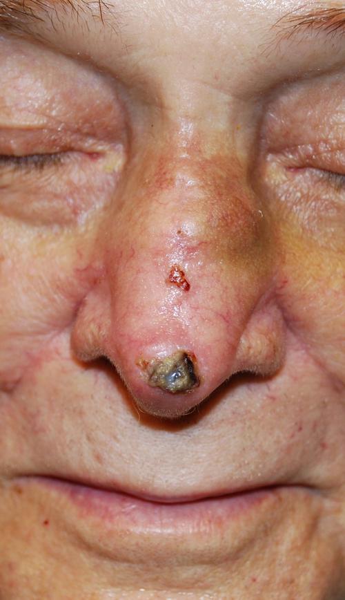
A 75 year-old male patient, on warfarin (blood thinner) therapy, presented to our office for a non-healing sore on his nose (Figure 1). According to the patient, the lesion had been treated with liquid nitrogen at least twice in the past 12 months. A biopsy demonstrated an infiltrative basal cell carcinoma. This type of basal cell carcinoma is typically more aggressive than a standard nodular basal cell carcinoma and is frequently larger than it appears to be. These cancers can cause severe disfigurement if left untreated. This cancer can grow both extremely wide and deep, causing it to destroy skin, deeper tissue, and can even damage bone. The most common place that a Basal Cell Carcinoma will present itself is on the nose.
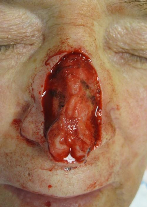
Three stages of Mohs micrographic surgery were required to clear the tumor. The final defect involved the majority of the nasal dorsum, nasal tip and a small amount of the bilateral nasal sidewall (Figure 2). Minor involvement of the nasal cartilage was noted.
Several closure options were discussed with the patient including but not limited to a paramedian forehead flap. This closure involves identifying the supratrochlear artery that supplies some of the skin on the forehead and dissecting the surrounding tissue to utilize for the repair. This is typically a two-stage process and can be difficult for some patients to tolerate, especially those on life saving blood thinners. Due to the complexity of this procedure, the patient wished to pursue other options.
Grafting was discussed, but he and his wife had significant concerns regarding the cosmetic results of a graft. Skin grafts can be very cosmetically pleasing when executed correctly. Unfortunately, in this case, the large defect size and exposure of cartilage would make the likelihood of a cosmetically pleasing graft very low.
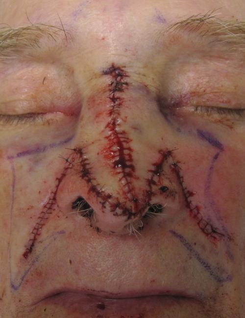
In the end we opted for a bilateral transposition flap. This flap had the advantage of being performed in a single stage while yielding a cosmetically pleasing result. Tissue from the middle part of each cheek was elevated and moved over to the intact tissue of the sides of the nose (transposed) and sewed into place over the defect (Figure 3).
The sutures were removed after one week of healing. At two weeks a small amount of scabbing is noted at the tip of the nose (Figure 4), but the patient and his wife were extremely happy with the result.
At Pine Belt Dermatology & Skin Cancer Center, we know that healthy skin is affected by more than just external care—it’s related to your overall health...
Here’s how UV light therapy works, why it is useful for scalp psoriasis during winter, what to expect from treatment, and how to use it safely.

A truly effective skincare routine should be tailored to your needs, protect your natural barrier, and target concerns with proven ingredients.
The cold, dry air outside combined with indoor heating can strip away your skin’s natural moisture, leaving it tight, flaky, and more vulnerable to irritation.

At Pine Belt Dermatology, we understand how winter weather affects your skin and how frustrating it can be to deal with the discomfort that comes with it.
Acne is often thought of as a summer skin concern. However, many people notice that their breakouts actually worsen in the fall.

This blog explores why SPF isn’t just a summer essential—it’s a daily requirement, no matter the season.
Fortunately, with the right approach and treatments, you can begin reversing these effects and restore your skin’s health and radiance.
Summer is a time for beach trips, backyard barbecues, and sunshine-filled adventures, but for many people, it also brings along an unwanted guest: acne.
Whether you're diving into a chlorinated pool or splashing in the salty waves of the beach, summer fun often comes with hidden consequences for your skin. While swimming is an excellent way to stay active and cool off, the effects of prolonged exposure to chlorine and saltwater can leave your skin dry, irritated, and vulnerable to damage.