
Mohs Surgery Case - Basal Cell Carcinoma on Eyelid & Discussion on Eye Tumors
Dr. David Roy • August 1, 2019


View more
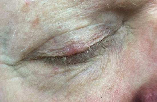
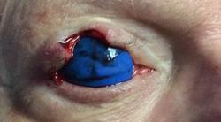
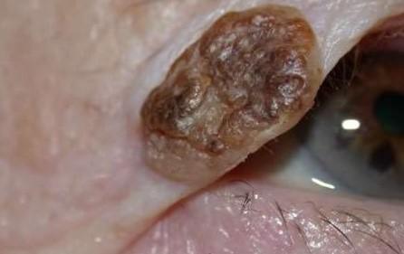
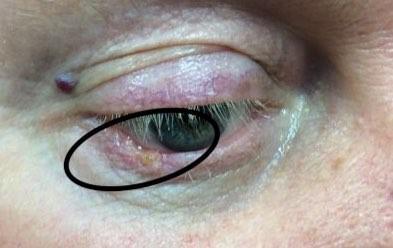
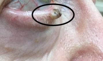
A 79 year-old male patient, presented to our office for the evaluation of a small growth on his upper eyelid (Figure 1). He stated that the spot had been present for approximately 1-2 years, but was unsure about the timeline. A biopsy of the lesion demonstrated a basal cell carcinoma. Two stages of Mohs micrographic surgery were required to clear the tumor. The final defect involved a significant portion of the patient’s upper eyelid and was full thickness in some areas (Figure 2).
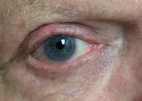
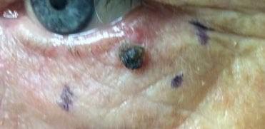
Several closure options were reviewed with the patient. He did not want to schedule a separate appointment with oculoplastic surgery and preferred to complete as soon as possible. Ultimately a simple wedge resection was performed to repair the defect (Figure 3).
Sutures were removed after one week and at follow up at 4 weeks, the patient demonstrated full function of the eyelid, with minimal scarring (Figure 4).
Further Discussion on Eyelid Tumors & Growths:
There are many things that can pop up on your eyelids. Some are harmless and some are not. The most common things that occur on the eyelids and prompt patients to see us for an evaluation are chalazions (styes), cysts, seborrheic keratoses, xanthelasma, syringomas, and hidrocystomas.
Chalazions
are also known as styes and are benign bumps caused by inflammation of various glands on the eyelids. They can be treated with both medication and at times with surgery. They can mimic things like basal cell carcinoma, so if you have a stye that is not improving, make sure that you have it evaluated by your physician.
Cysts
are benign growths that can occur from different structures in the skin. Which structure they develop from defines what type of cyst you have. They can be commonly found on the eyelids and can be derived from glandular tissue, skin, or hair follicles. “Sebaceous cyst” is a generic term that is used frequently to describe a number of different cysts that have a white, cheesy material in them. It is not a specific term that defines any one cyst.
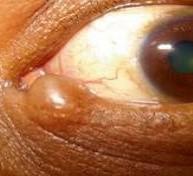
Hidrocystomas
are small cystic growths that derive from sweat glands or other specialized glands in the eyelid. They present as small smooth bumps and are easily mistaken for basal cell carcinoma. Some will have a faint blueish color to them. When pooped, a clear, water-like liquid will drip from them. This is sweat!
Seborrheic keratoses
are one of the most common growths found on the skin and they can occur on the skin of the eyelids. They are typically raised, rough and can range from skin colored to dark black. Moles and even melanoma can look similar to these, so it is very important to have them evaluated by a health care professional.
Moles.
The fancy name for a mole is a melanocytic nevus. These can occur anywhere on your body and the eyelid is no exception. They can even develop on certain tissues in your eye. While most moles are benign, it is very important to watch them for change and to have them evaluated on a regular basis.
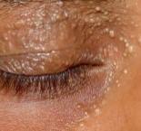
Syringomas
are small white bumps that appear, typically around the eyelids. They are completely benign yet are often of great cosmetic concern to patients. Syngomas are derived from sweat glands and are distinct from the hidrocystomas that we discussed earlier. They can be few or numerous and can be treated easily. Unfortunately, patients will typically develop new lesions after treatment.
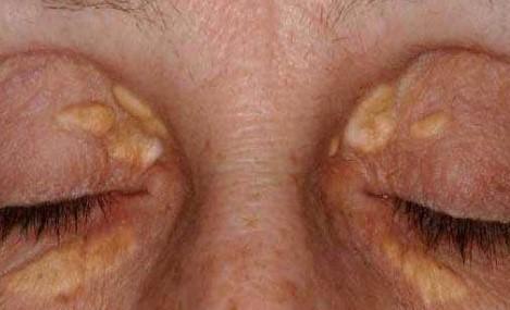
Xanthelasmata
are deposits of fats that have been picked up by specific cells in our skin and show up as white to yellow bumps or patches on our eyelids. Most patients with these lesions on their eyelids have normal levels of fats in their blood, however on rare occasions, this can be a marker for abnormal elevations of these fats and even a sign of genetic abnormalities that can be passed on to your children.
Cancer can develop on the eyelid. These include basal cell carcinoma, squamous cell carcinoma, sebaceous carcinoma, malignant melanoma, merkel cell carcinoma, lymphoma, and metastatic tumors from cancers in other parts of the body. We will briefly review a few of the more common types.
Basal cell carcinoma
is not uncommonly found on the eyelid and is often seen on the lower eyelid. While it is a slow growing tumor, it can be very locally destructive and can invade the tissue on the eye, behind the eye, or even involve the muscle and bone that surrounds the eye. In extreme and neglected cases it can metastasize, but this is very rare.
Squamous cell carcinoma.
This is the second most common skin cancer and it is the most common non-melanoma skin cancer seen in skin of color. These lesions can pop up out of nowhere, or they can develop from precancerous lesions known as actinic keratoses.
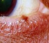
Sebaceous carcinoma
is a less common tumor derived from the oil glands found on the skin. The eyelid is one of the most common places for these cancers to develop. The tumor seen in the photo below is a very subtle red bump. What prompted the patient to seek attention was the small scab that kept forming on the lesion. This tumor can spread, so surgery is very important in order to clear the tumor and hopefully prevent spread.
Malignant melanoma
can occur anywhere on the body and can be seen on the eyelid and even on and within the eye itself. It can develop from a mole or it can develop out of the blue. Prompt evaluation is essential so if you see a dark spot that is new or changing, make sure you have it evaluated.

At Pine Belt Dermatology & Skin Cancer Center, we know that healthy skin is affected by more than just external care—it’s related to your overall health...

Here’s how UV light therapy works, why it is useful for scalp psoriasis during winter, what to expect from treatment, and how to use it safely.

A truly effective skincare routine should be tailored to your needs, protect your natural barrier, and target concerns with proven ingredients.
The cold, dry air outside combined with indoor heating can strip away your skin’s natural moisture, leaving it tight, flaky, and more vulnerable to irritation.

At Pine Belt Dermatology, we understand how winter weather affects your skin and how frustrating it can be to deal with the discomfort that comes with it.
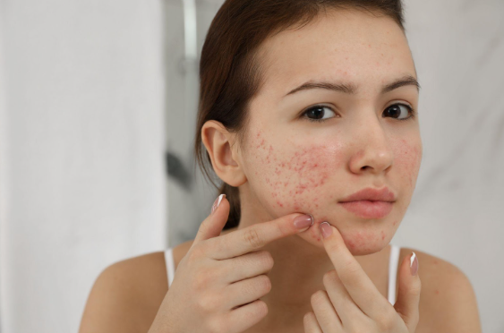
Acne is often thought of as a summer skin concern. However, many people notice that their breakouts actually worsen in the fall.

This blog explores why SPF isn’t just a summer essential—it’s a daily requirement, no matter the season.
Fortunately, with the right approach and treatments, you can begin reversing these effects and restore your skin’s health and radiance.
Summer is a time for beach trips, backyard barbecues, and sunshine-filled adventures, but for many people, it also brings along an unwanted guest: acne.
Whether you're diving into a chlorinated pool or splashing in the salty waves of the beach, summer fun often comes with hidden consequences for your skin. While swimming is an excellent way to stay active and cool off, the effects of prolonged exposure to chlorine and saltwater can leave your skin dry, irritated, and vulnerable to damage.