Case of the Month February 2020
February 17, 2020
My fourth year of medical school, I was on a dermatology rotation and I had the pleasure of helping care for a female patient with a squamous cell carcinoma of her left index finger. She was sent to the hand surgeon for treatment and two weeks later she returned to the office for follow up. I was shocked to see that her finger had been amputated. It was completely gone! At the time I realized that although I had not thought about it prior, this was a high-risk tumor in a high-risk area and removing the finger was probably the best mode of treatment to ensure that the tumor was completely removed and to prevent metastasis.
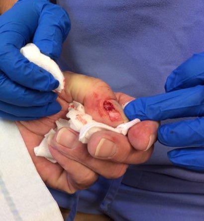
Fast forward 14 years. A 71-year-old male patient presented to our office for the evaluation of a painful lesion under his right thumbnail. He had been treated for a fungal infection of the nail for the last 12 months with no improvement and he was seeking a second opinion. The nail plate and cuticle were elevated revealing an erythematous tumor (Figure 1). A biopsy of the lesion demonstrated a well-differentiated squamous cell carcinoma. Squamous cell carcinomas involving this anatomical location are considered high-risk tumors and have the potential for metastasis.
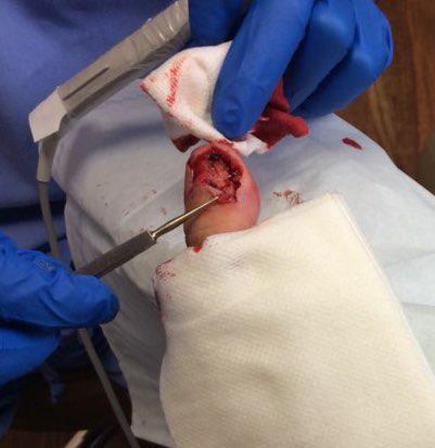
Due to the aggressive nature of the lesion and in an attempt to save the thumb from amputation, Mohs surgery was performed. After 3 stages of surgery the final defect can be seen below (Figure 2).
Partial removal of the nail matrix (the portion of the nail that creates new cells and allows the nail to grow) was required to clear the tumor. This results in permanent loss of that portion of the nail.
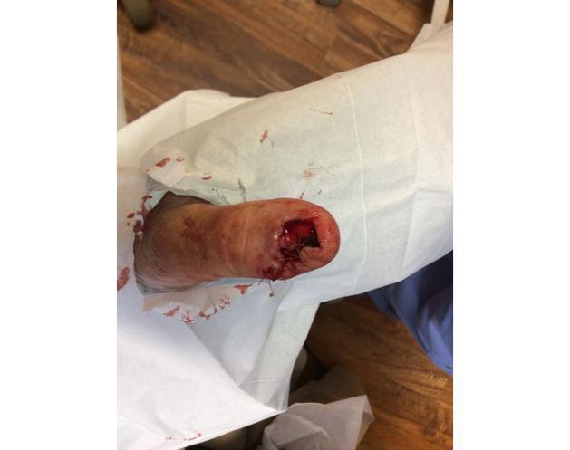
Closure options for this defect included a skin graft, secondary intention healing, and local tissue flaps. The patient stated that he did not want a graft and he did not want to let it close on its own. Together with the patient and his family, we agreed upon a laterally based rotation flap. The final closure can be seen below (Figure 3).
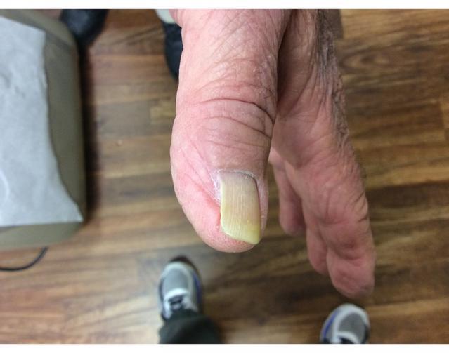
Sutures were removed after two weeks at which time excellent early surgical results were noted. Normal anatomy was maintained with the exception of the partial nail loss. Overall, the thumb looked anatomically correct and was fully functional. He and his family were very happy with the results. At four years, no evidence of recurrence is noted, and the patient has a fully functioning and normal looking thumb (Figure 4).
Your skin, the body's largest organ, is a vital indicator of your overall health. Changes in its appearance can signal underlying medical conditions that may require attention. Whether it’s ensuring you’re getting the right nutrients, managing stress, or seeking professional care, being attuned to what your skin tells you makes it easier to stay on top of your health.
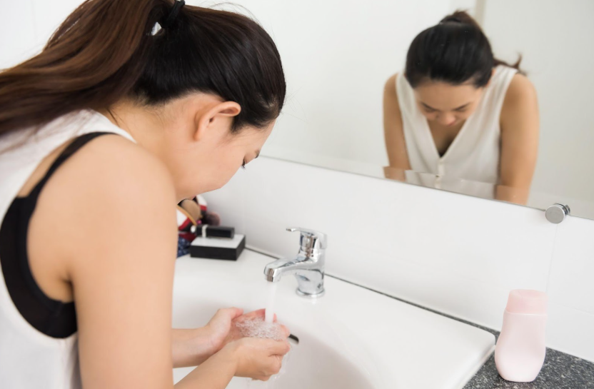
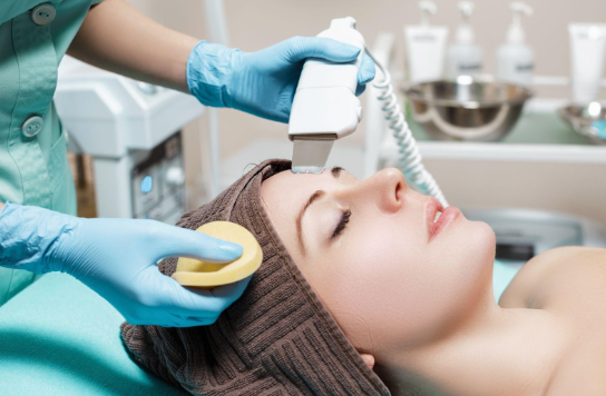
Achieving healthy, glowing skin begins with an effective cleansing routine. However, small mistakes can disrupt your skin’s natural balance, leading to irritation, breakouts, or premature aging. Avoiding these common errors and incorporating advanced techniques can significantly impact your skincare journey.

You’ve likely felt it in your life: that uncomfortable feeling of tightness, flakiness, and sometimes even itchiness that can make your skin look and feel less than its best. But what exactly causes dry skin, and how do you treat it? Don’t fret because we’ve put together some treatment tips that can help your dry skin regain its moisture.

