Mohs Case - Recurring Basal Cell Carcinoma In Young Patient
Dr. David Roy • December 9, 2019
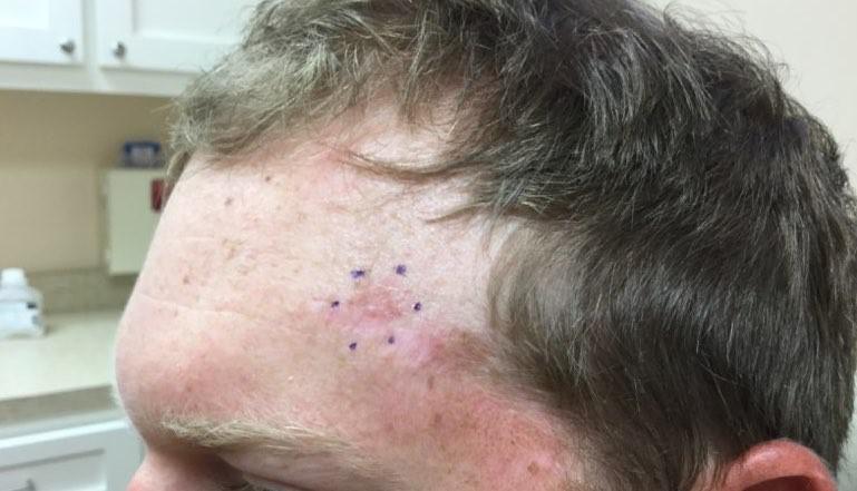
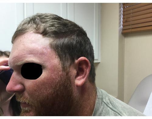
A 27-year-old male presented to our office for the treatment of a recurrent infiltrative basal cell carcinoma of his left forehead (Figure 1). According to the patient, surgery had been attempted twice previously. Neither attempt yielded clear margins and the tumor recurred. As noted in previous blogs, this type of basal cell carcinoma is typically more aggressive than a standard nodular basal cell carcinoma and is frequently larger than it appears to be. This is why it is difficult to obtain clear margins with standard surgery (wide excision).
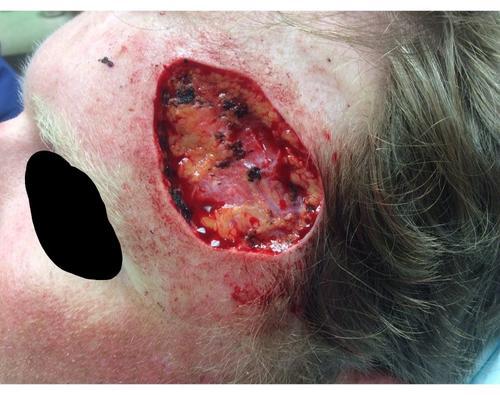
Five stages of Mohs micrographic surgery were required to clear the tumor. The final defect involved a significant portion of the patient’s forehead (Figure 2).
As you can see, there was a significant difference between what appeared to be the size of the lesion before surgery, and the actual clear margins following Mohs.
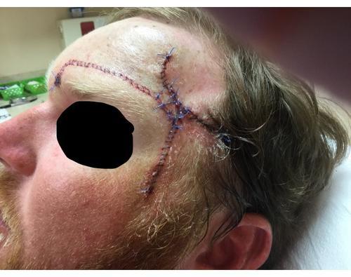
Several closure options were reviewed with the patient. Grafting was discussed, but I had significant concerns regarding the look of a graft given his age and the location of the defect. Ultimately, the patient opted for a combination advancement/rotation flap. This flap had the advantage of being performed in a single stage while yielding a relatively pleasing cosmetic result. Tissue from the medial aspect of the forehead was elevated and moved (advanced) centrally. The skin of the patient’s left temple was also mobilized and rotated upward. This allowed for complete closure of the wound while keeping the patient’s left eyebrow level (Figure 3).
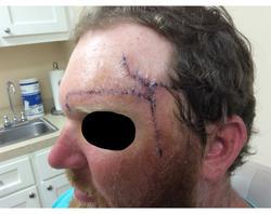
The sutures were removed after one week of healing (Figure 4 a-b). The patient was extremely happy with the results and reported no complications.
At his two-year follow-up, there is no evidence of recurrence and an acceptable cosmetic result has been achieved (Figure 5).

Your skin, the body's largest organ, is a vital indicator of your overall health. Changes in its appearance can signal underlying medical conditions that may require attention. Whether it’s ensuring you’re getting the right nutrients, managing stress, or seeking professional care, being attuned to what your skin tells you makes it easier to stay on top of your health.

Achieving healthy, glowing skin begins with an effective cleansing routine. However, small mistakes can disrupt your skin’s natural balance, leading to irritation, breakouts, or premature aging. Avoiding these common errors and incorporating advanced techniques can significantly impact your skincare journey.

You’ve likely felt it in your life: that uncomfortable feeling of tightness, flakiness, and sometimes even itchiness that can make your skin look and feel less than its best. But what exactly causes dry skin, and how do you treat it? Don’t fret because we’ve put together some treatment tips that can help your dry skin regain its moisture.
